Basic anatomy & Radiology for breast cancer case - Download as a PDF or view online for free This document provides an overview of the anatomy relevant to breast cancer case delineation. It describes the layers of the chest wall including skin, fat, muscles and bones. It outlines the anatomy of structures in the chest including the sternum, ribs, vertebrae, shoulder girdle, and vessels in the neck and chest. The document also details the anatomy of the breast, axilla, supraclavicular fossa, and various muscles of the chest, back, neck and shoulder including the pectoralis major, deltoid, trapezius, and sternocleidomastoid.
Invasive Ductal Carcinoma (IDC)
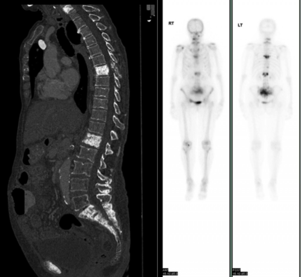
AuntMinnie.com - A 48-year-old woman with a history of breast cancer presented for a bone scan. A routine bone scan was performed. Can you solve this #radiology case from Dr. David Tischfield
Atlas of breast cancer early detection
Basic anatomy & Radiology for breast cancer case
Ductal carcinoma in situ, Radiology Reference Article
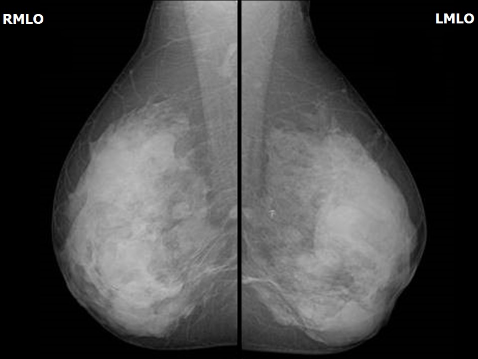
D&C exam 1 Mammography and DEXA Flashcards
Bone metastases - CT and bone scan - Radiology at St. Vincent's University Hospital
Atlas of breast cancer early detection
A case with metastatic breast cancer at the sternum. (A) ¹⁸F-FDG-PET
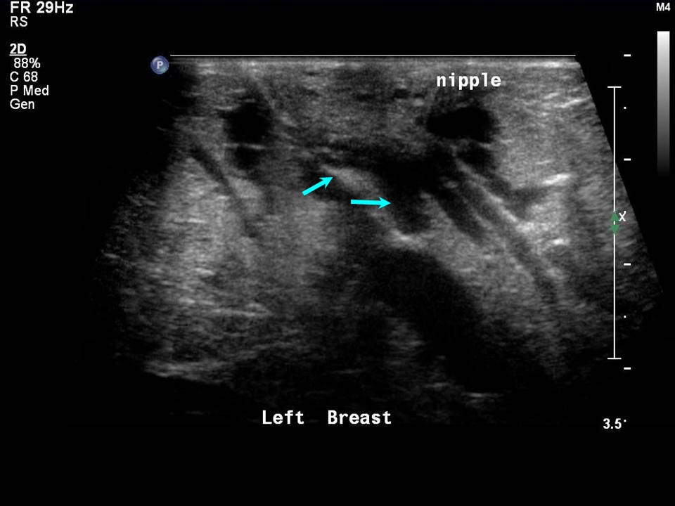
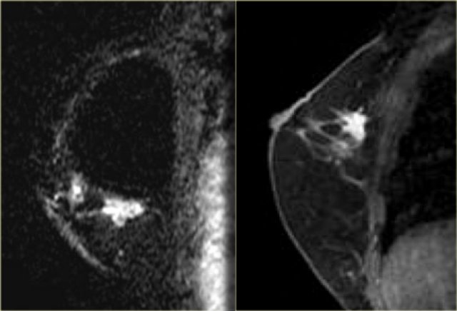
The Radiology Assistant : MRI of the Breast
ProFound AI Breast Cancer Health Suite
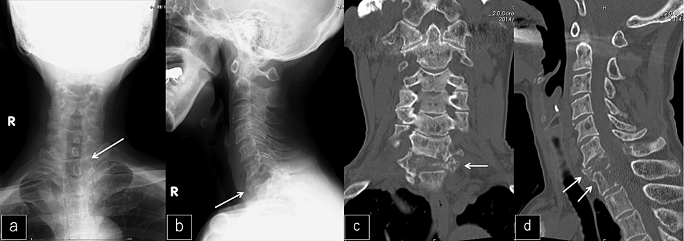
Cureus, The Decalcification of Cervicothoracic Spinal Metastasis of Breast Cancer Due to Discontinuation of Denosumab: A Case Report
Clinicopathologic and mutational profiles of primary breast diffuse large B cell lymphoma in a male patient: case report and literature review, World Journal of Surgical Oncology
Squamous cell carcinoma of the breast: About a case - ScienceDirect