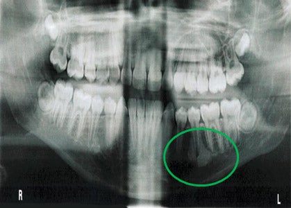
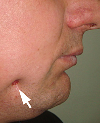
a Mandibular fistula indicated by an arrow in the apical region of dd
$ 33.50
4.6(550)In stock
Download scientific diagram | a Mandibular fistula indicated by an arrow in the apical region of dd 36-37. b A fistula in the apical region of dd 46-47 (white arrows) and a red area in the mucosa (black arrows) are seen in the right lingual surface of the mandible. c Panoramic radiograph showing no bone lesions in the mandible. d Periapical x-ray with no bone involvement in the apical region of dd 46-47 from publication: Treatment of bisphosphonate-induced osteonecrosis of the jaws with Nd:YAG laser biostimulation | Osteonecrosis, Jaw and Nd:YAG Laser | ResearchGate, the professional network for scientists.
Frs hfn
Satu ALALUUSUA, University of Helsinki, Helsinki, HY, Institute of Dentistry
Case Archive, School of Dental Medicine
/profile/Maria-De-Souza-3/publ
Satu ALALUUSUA, University of Helsinki, Helsinki, HY, Institute of Dentistry
a Mandibular fistula indicated by an arrow in the apical region of dd
Case Archive, School of Dental Medicine
Benign Lumps and Bumps
Satu ALALUUSUA, University of Helsinki, Helsinki, HY, Institute of Dentistry
Single and Multiple Odontogenic Cutaneous Sinus Tracts
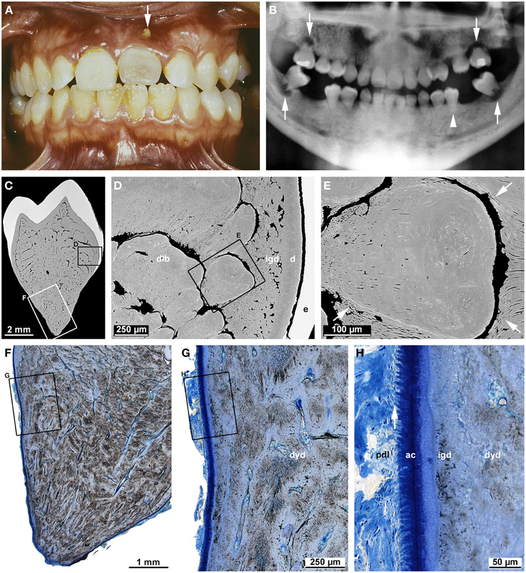
Frontiers Malformations of the tooth root in humans
Retreatments Solutions For Periapical Diseases of Endodontic Origin, PDF, Dentistry